Muscle Swimmer
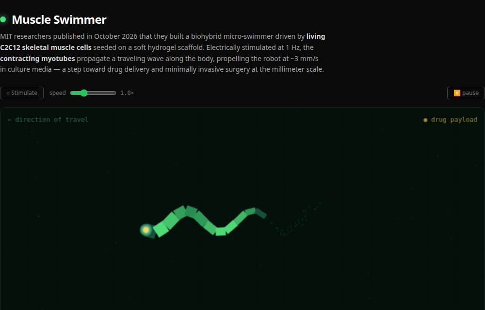
MIT researchers published in October 2026 that they built a biohybrid micro-swimmer…
Full description
MIT researchers published in October 2026 that they built a biohybrid micro-swimmer driven by living C2C12 skeletal muscle cells seeded on a soft polyethylene glycol diacrylate (PEGDA) hydrogel scaffold — C2C12 myoblasts are cultured on a fibronectin-coated PDMS substrate, differentiated into multi-nucleated myotubes in a low-serum medium, aligned along the direction of thrust by 1–5 µm micro-grooves etched into the surface, then bonded to the 3D-printed robot body and electrically stimulated at 1 Hz with a 2.5 V/cm field that drives action potentials in every myotube simultaneously, producing a synchronous contractile wave that propagates head-to-tail and propels the robot at ~3 mm/s in culture media; watch the soft segmented swimmer undulate through fluid with a yellow drug payload at its head and animated wake particles streaming behind, toggle electrical stimulation to increase wave amplitude by 40% and boost swimming speed as blue lightning arcs fire across the body, drag the speed slider from 0.3× to 3× to control tempo, pause the animation by clicking the canvas, browse eight fact cards covering C2C12 cell culture and myotube differentiation, the Stokes-flow physics of micro-scale undulatory swimming, why living muscle outperforms synthetic actuators below 1 cm, the microfluidic oxygenation channels that extend cell viability to >30 days, magnetic steering for targeted drug delivery, channelrhodopsin-2 optical control as a wireless future direction, and the scaling-law argument that favors biohybrid designs at millimeter scale, and trace the full C2C12 myoblasts → fibronectin PDMS scaffold → myotube differentiation → micro-groove alignment → PEGDA hydrogel body → 1 Hz electrical stimulation → synchronous contraction wave → undulatory swimming ~3 mm/s → drug payload delivery → minimally invasive surgery pipeline in a Mermaid flowchart.